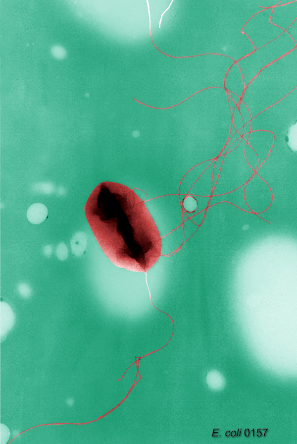
10) polysomes or poly-ribosomesThe outer cell membrane or outer membrane (OM) consists of a lipid bilayer structure composed of an outer layer or leaflet consisting of lipopolysaccharide (LPS) and an inner leaflet consisting of phospholipids. LPS is composed of 3 components: lipid A, a branched sugar chain and the O-antigen. Lipid A is made of 2 glucosamines attached to phosphates and linked to C14 3-hydroxy myristic acid. The branched sugar consists of two types of sugars, one a heptose and the other keto-deoxyoctonoic acid. The O-antigen consists of a long (up to 40 sugars) carbohydrate chain. Each LPS unit is covalently linked to form a trimer through pyrophosphate linkages to the sugars of lipid A. LPS is highly immunogenic and frequently toxic (i.e. E. coli O157). The other major components of the outer membrane are proteins -- largely consisting of porins (approximately 60,000) which coexist with LPS. The outer membrane is a barrier that is quite resistant to chemicals and hydrophobic compounds, including antibiotics. Porins are passive diffusion channels that allow hydrophilic molecules (i.e. nutrients) of up to 800 daltons to pass through. The width of the outer membrane is about 10-15 nm.The cell wall, which lies just below the outer membrane is composed of peptidoglycan (also known as murein or Braun's lipoprotein) which, in turn, is covalently bound to the outer membrane. The cell wall prevents the cell from being osmotically lysed and gives the cell its characteristic shape. It is technically a single supermolecule. Peptidoglycan (PG) is a loosely (30%) cross-linked polymer consisting of covalently linked sugar and peptide units. The sugar units are N-acetylgulocsamine and N-acetylmuramic acid. The peptides are tetrapeptides consisting of L-Ala-D-Gly-DAP-D-Ala, where DAP is diaminopimelic acid. Peptidoglycan is synthesized via the insertion of rings (about 1100 in all) that grow from about 200 different locations around the cell (which translates to 250,000 copies of murein). The spacing of these PG growth rings is about 1.3 nm. In E. coli the peptidoglycan layer is very thin and may only be a monolayer.The inner membrane is composed of a lipid bilayer about 8 nm thick consisting of ~40% phospholipids and 60% protein. The phospholipids include phosphatidylethanolamine (75%), phosphatidylglycerol (18%), cardiolipin (5%) and phosphatidylyserine (2%). The lipid (fatty acid) chains are mostly C16 palmitic acid (43%), C16 palmitoleic acid (33%) and C18 vaccenic acid (24%), which form ester links to create the phospholipids. There are no sterols or steroids in the inner membrane of bacteria. Recall that mesophiles (like E. coli) tend to have fatty acids with shorter chains and more unsaturated fatty acids to maintain membrane fluidity. The inner membrane, in combination with the outer membrane (i.e. the cellular envelope) serves as an osmotic barrier, a nutrient-specific transporter, a lipid synthesizer, a peptidoglycan synthesizer, electron transport system, a place for assembly and secretion of envelope proteins, a mechanism for chromosomal segregation and a site for chemo-sensing. There exist a number of regions (~200 per cell) called Bayer's junctions where the inner (cytoplasmic) membrane contacts the outer membrane. It is not known what these junctions do.The periplasm, which is about 10 nm thick, occupies between 10 and 20 percent of the volume of an E. coli cell. It is the space between the inner and outer membrane and houses both proteins and the cell wall (peptidoglycan). It is thought to help in osmoregulation. The periplasm contains a number of proteins essential for nutrient binding, degradative enzymes (proteases, endonucleases), detoxifying enzymes (beta lactamase), peptidoglycan synthesis, cytochromes (electron transport) and chemotaxis or chemosensing proteins. The periplasm contains approximately 80,000 proteins.On the surface of the outer membrane can be found flagella. Flagella are rigid screw-like appendages (10-20 microns in length and approximately 25 nm wide) anchored to the outer membrane that rotate (clockwise or counterclockwise) in a propeller like fashion to facilitate bacterial movement. When flagella rotate counterclockwise this creates a pushing force that allows the bacterium to move (called a run). When flagella rotate clockwise the bacterium tumbles or twiddles. The change between a run and a twiddle is brought about by subtle changes to the structure of flagellar filament proteins (flagellin of FliC). Each flagellar filament is composed of 11 protofilaments wound in a bundled helix composed purely of flagellin. The orientation of these protofilaments (and the structure of the whole flagellar filament) is affected by small cumulative changes in the flagellin monomers brought on by chemo/osmotactic forces. Flagella are remarkably complex motor systems consisting of up to 50 different proteins which spontaneously self-assemble to form nano-scale rotors, stators and power (ATP) supplies. There are three components to a flagellum: the filament (composed of 30,000-40,000 flagellin monomers), the hook (which differ between G+ and G- cells) and the basal body (motor and power supply). A typical bacterium may have from 5-20 flagella. In E. coli, the flagellar are arranged in a peritrichous fashion (meaning they are scattered uniformly around the cell).Fimbrae or pili are thin appendages commonly found on G- cells. They are approximately 6.5 nm in diameter and between 200 and 2000 nm in length, meaning that they are smaller than flagella. A cell may have from 100-300 pili or fimbrae, meaning that they are much more numerous. The primary component in pili is the papA protein, a 16 kD protein which self-assembles into a helical repeat creating a hollow (1.5 nm) core of superstructured protein. Approximately 300 (short) to 3000 (long) papA proteins are needed to make a single pilus. The term fimbrae is used when referring to filaments responsible for surface attachment. The term pili refers to filaments used to mediate attachment to other bacteria (bacterial conjugation or DNA transfer).Also on the surface (i.e. outside the outer membrane) of E. coli are crystalline-like surface proteins which self-assemble to form S-layers. These S proteins form complex, rigid polyprotein networks that may be important for cellular protection and adherence.The cytoplasm is where all other major components of an E. coli cell reside. The cytoplasm contains the chromosomal DNA (about 2.3 genomes worth in an actively dividing cell), the RNA (tRNA, mRNA and rRNA), the ribosomes or polyribosomes (for protein synthesis), inclusion bodies or storage granules, essential ions (120 million), small organic molecules (18 million) and about 2.1 million proteins. The region of the cytoplasm containing the chromosome is called the nucleoid. It contains up to 2 chromosomal equivalents (in rapidly dividing cells). Each chromosome measures 1.55 mm in circumference (490 microns in diameter). The chromosome interacts with tens of thousands of nuclear proteins (HU, IHF, H-NS) with their being sufficient HU protein to bind the DNA every 200 bp (40,000+ monomers for each chromosome, HU dimerizes). These pseudo-histones condense the chromosome into a body (called a nucleiod) about 17 microns in diameter. The prokaryotic ribosome is composed of 2 subunits, the 30S and the 50S subunits. The 20S subunit has 21 proteins bound to the 16S (1700 nt) rRNA. The 50S subunit has 34 proteins bound to the 23S (3700 nt) and 5s (120 nt) rRNA. The storage granules or inclusions include metachromatic granules (which contain polyphosphate), glycogen granules (which store polyglucose) and lipid inclusions (which contain poly-B-hydroxybutyrate or PHB). Bacteria do not have a nucleus or other complex organelles (such as mitochondria, endoplasmic reticulum or chloroplasts) as found in eukaryotic cells. This simple interior structure makes bacterial cells much simpler to model and much easier to understand than eukaryotic cells. (From http://redpoll.pharmacy.ualberta.ca/CCDB/intron_new.html) (BacMap)